
 |
 |
| Senior Staff Koiku Asakura, M.D. |
“Diagnostic Radiology” is to examine organs, which are not visible directly from outside the body, by creating radiological images on various modalities. They include CT (computer tomography) scanning using X-ray, and MRI (magnetic resonance imaging), which is not radiological but using magnetic energy. They make it possible to detect tumors at early stages, to assess how deep and wide they are spreading, and to make certain if there are recurrences or metastases after cancer treatments. Therefore, diagnostic radiology plays a crucial role for optimal cancer treatments.
At the Division of Diagnostic Radiology, we accept orders for image examinations (including general X-ray examination, gastroenterological contrast imaging, CT, MRI and angiography) from other clinical departments and provide them with images and diagnostic reports.
* Cancer treatments provided using imaging modalities are basically handled at the Division of Interventional Radiology (IVR).
CT scanning or MRI is essential for diagnosing a cancer. Lately, due to the advancement of technologies, new models for CT and MRI equipped with newly-developed functions have come out, which have made it possible to obtain more precise image information than ever. At the SCC, these new models have been installed and been widely used for cancer diagnoses. For example, the latest CT scanner can collect very fine image data while scanning at a very high speed, and create multiplanar images including sagittal-sectional images and three-dimensional images not limited to just coronal-sectional ones, with its multi-detector functions. Furthermore, we have two new integrated PET/CT machines, which enable PET (positron-emission tomography) examination for detecting cancers in the entire body all at once combined with CT scanning. They are fully in operation now, helping us diagnose cancers at the early stages, assess the treatment effects, and detect recurrences or metastases as early and precise as possible.
The number of cases for diagnostic imaging is about 110,000 a year here, including those done with general X-ray and the others done at highly-advanced new modalities. The bigger the number is, the more opportunities to enhance skills and expertise for image-taking and the interpretation. It has made us utilize the utmost merits of highly-advanced modalities. That way, we can provide fine image data and very reliable diagnostic reports based on them.
We accept orders for image examinations from other medical institutions in the eastern region of Shizuoka Pref. and provide them with the diagnostic reports, in accordance with the medical collaborative alliance structure consolidated for efficiently utilizing the costly advanced modalities.
At the Shizuoka Cancer Center, the latest, cutting-edge modalities for diagnostic images have been installed. We utilize their utmost functions effectively, doing our best to let many patients take the image examinations as promptly as possible. We also try to come up with precise and reliable diagnoses, while at the same time, we try to utilize the utmost merits of the modalities and our own acquired skills and expertise. Practicing and developing minimally invasive image-guided treatments are what we aim for at this division.
At the SCC, what is done at the medical examination section in the department of radiology in most big hospitals is divided into three divisions; the Divisions of Diagnostic Radiology, IVR and Radiation Therapy. For cancer diagnoses, the Divisions of Diagnostic Radiology and IVR work tightly together, making collaborative efforts to contribute to the SCC Hospital providing precise and reliable diagnoses and effective treatments.
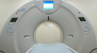
Diagnostic imaging includes plain radiography, fluoroscopy, bone mineral quantum analysis, CT, MRI, angiography, and nuclear medicine. Among all the modalities for imaging we have, it is noteworthy that we have 4 advanced multi-detector CT scanners. Two of them are high-definition CT scanners having twice as high spatial resolution capability for in-plane and body-axis directions each. The other two are such cutting-edge models as having 320-row detectors so-called aero CT. With them, we handle 150 cases a day in average, which makes about 35,600 cases a year. CT examination is done with a diagnostic equipment consisting of X-ray tubes (devices to deliver X-ray) spinning around a lying patient for scanning, detectors to receive scanned data, and a computer to analyze them and create images based on them. The latest CT makes it possible to scan at a high speed and to collect very fine image data at the same time. The images created from the obtained data can be multiplanar including lengthwise-sectional or three-dimensional, not just limited to cross-sectional. They play critical roles in cancer medicine, as they offer information essential for all the cancer diagnoses, staging, pre-operative vascular anatomy and decision-making for the extent of resection.
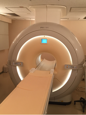
For MRI (magnetic resonance imaging), we have 3 cutting-edge models including two 3T (tesla)-models and one 1.5T-model. With 3 of them, we handle 45 examinations a day in average, which makes about 11,100 cases a year. MRI machines use strong magnetic fields and radio waves to take cross-sectional images from various angles of a human body. Especially for the brain, the spine, four limbs and interior of the pelvis where the movements are very well-controlled during the examination, we can get very clear-cut images. MRI offers critical information for cancer diagnoses just like CT, with no radiation exposures which is a huge merit. However, there are some demerits as well, including longer examination time than CT, big noises, and making a patient stay in an enclosed space which makes it hard for those with claustrophobia. In addition, when a patient has a metallic object implanted, such as a heart pacer, there will be no way to take an examination with MRI. Yet, it is quite useful for diagnosing brain tumor and breast cancer especially, as the 3T-MRI offers images with higher resolutions than the 1.5T model due to the twice-as-high magnetic field intensity. Naturally, it is useful for diagnosing not only malignant tumors but acute-staged cerebral infarctions.
Furthermore, we handle nuclear medicine examination using radioactive tracer. Nuclear medicine examination is a diagnostic method with radioactive tracer administered, of which disposition is examined externally to assess the pathological condition. Imaging is usually done with gamma camera, SPECT (single photon emission computed tomography) and PET (positron emission tomography). At the SCC, PET is the main diagnostic examination for cancer. With a medical small cyclotron installed in the hospital, a radioactive glucose mimicker called 18F-FDG (18F fluorodeoxyglucose), which resembles glucose, is synthesized for PET examination. As cancerous cells consume more glucose than normal cells to gain energy for proliferation, 18F-FDG is absorbed and accumulated more in them just like glucose than normal cells. PET examination utilizes such abnormal accumulation of radioactive agent for imaging, which can be quite helpful for cancer diagnoses at early stages, efficacy assessment of treatments, and detecting recurrences or metastases.
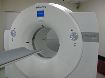
We have two units of PET/CT (equipment combining PET with CT), one of which is a quite-advanced, state-of-the-art model equipped with a variable-speed, continuously-moving scanner. It has shortened the examination time dramatically. Due to the speed, we handle 20 cases a day in average with PET/CT, which makes about 3,800 cases a year.
PET/CT imaging test can be covered by the Japanese public health insurances for diagnosis for malignant tumor excluding early-staged gastric cancer, but there are still some difficult cases of which diagnoses can’t be ensured even with this method. Please ask your primary doctor for clarification when you have an inquiry about the examination.
We also handle bone scintigraphy tests for diagnosing bone metastases, and sentinel lymph node scintigraphy tests for diagnosing metastases in lymph nodes at this Division.
diagnostic imaging, specifically for the chest area
diagnosis and treatment of malignant tumor in the chest
IVR procedures for the respiratory organs
diagnosis of drug-induced pulmonary disease
Diagnostic Radiologist Certified by the Japan Radiological Society
Certified Specialist and Supervisory Doctor, Councilor, the Japan Society for Respiratory Endoscopy
Certified Specialist and Certified Doctor for PET Nuclear Medicine, the Japanese Society of Nuclear Medicine
Councilor, the Japan Lung Cancer Society
The Japanese Society of Interventional Radiology
Doctor for Radiogram Interpretation at the Breast Cancer Screening Certified by the Japan Central Organization on Quality Assurance of Breast Cancer Screening
Educational Committee Member, the Japanese Board of Cancer Therapy
Visiting Professor, Fujita Health University School of Medicine
diagnostic imaging in general
Diagnostic Radiologist Certified by the Japan Radiological Society
Certified Specialist and Certified Doctor for PET/Nuclear Medicine, the Japanese Society of Nuclear Medicine